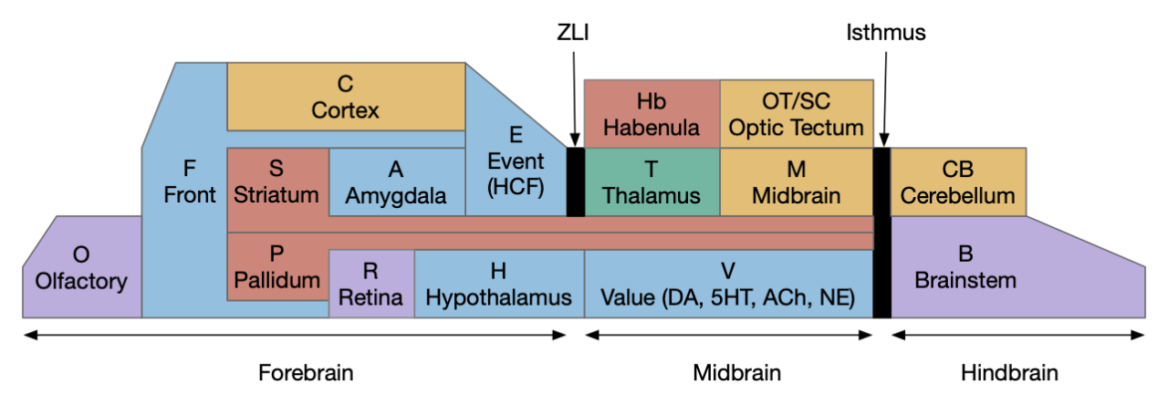
Previous essay 23 (feeding and neuropeptide core) and essay 27 (feeding state machine) covered feeding, but were not based on the ascidian tunicate brain. After studying tunicates in essay 30 (RTPA – real time place avoidance), I think using the adult ascidian brain is a better foundation. The earlier essays chose the center of feeding at H.l (lateral hypothalamus), which is a foraging locomotion center, but this essay centers feeding around eating in R.pb (parabrachial). Where R.pb is centered around eating, tasting, and digestion, H.l is centered of seeking and locomotion. Because adult ascidians are sessile, they have no locomotion, but they do eat by filter feeding, suggesting that the eating functions in R.pb are more fundamental to the feeding process than the locomotive foraging.
Adult ascidian
The adult ascidian tunicate (specifically Ciona “sea squirt”) is a sessile filter feeder. Like other chordates, ascidian filter feeding uses pharyngeal slits, which developed into gills for vertebrates, and later into the jaw. The ascidian larva phase is only 24 hours and can swim like vertebrates, using phototaxis and geotaxis to find an appropriate permanent settling place. The adult ascidian brain dissolves the larva navigation areas and expends the “neck” of the larva brain, between the sensory ganglia and the motor ganglia [Gigante et al 2023].

The adult ascidian brain is marked by the Phox2 genetic transcription factor [Gigante et al 2023], which corresponds to specific vertebrate hindbrain areas. In vertebrates Phox2b corresponds to much of the hindbrain, including N5 (trigeminal), N7 (facial), N10 (vagus), N11 (accessory) nerves, R.nts (nucleus of the solitary tract), and R.na (nucleus ambiguous). The vertebrate Phox2a corresponds to V.lc (locus coeruleus – norepinephrine), N3 (oculomotor), and N4 (trochlear) nerves [Dufour et al 2006]. To put it another way, the branchial (pharyngeal) motor neurons are a distinct group consisting of N5, N7, N9 (glossopharyngeal), N10, and N11 [Fritzsch et al 2017].
The Phox2b nuclei essentially match the parasympathetic system:
- N5 – trigeminal – jaw, chewing, face and mouth
- N7 – facial – facial expressions and taste in tongue
- N9 – glossopharyngeal – taste in tongue, swallowing, blood pressure
- N10 – vagus – heart, breathing, digestion, etc.
- N11 – accessory – shoulder and neck
- R.nts – nucleus of the solitary tract – visceral and taste sensorimotor
- R.na – nucleus ambiguous – speech, swallowing, cardiac
Note that the jaw develops from the first pharyngeal arch, and the pectoral girdle (shoulder and neck) may develop from the last pharyngeal arch [Brazeau et al 2023].
The Phox2a nuclei are:
- N3 – oculomotor – most eye movement
- N4 – trochlear – additional eye movement
- V.lc – locus coeruleus – main source of norepinephrine
Because the ascidian adult brain is necessarily self-contained, corresponding to a restricted set of hindbrain ganglia, it’s a good center module for vertebrates, as a thought experiment. However, it’s important to remember that tunicates are highly divergent from the original vertebrate/tunicate ancestor and trying to derive that ancestor from ascidians is extremely suspect [Holland 2015].
R.pb parabrachial nucleus
For the sake of the simulation, I’m combining R.pb with associated nuclei like R.nts, R.na, and R.plc (pre locus coeruleus). Science research needs to distinguish the nuclei, of course, but complicating the essay’s model wouldn’t add much verisimilitude.
R.pb is a highly heterogenous area [Pauli et al 2022] with at least 36 gene expression clusters [Nardone et al 2024]. Its neurons derive from two distinct progenitor populations marked by transcription factors lmx1b and foxp2 [Karthik et al 2022]. The lmx1b population projects to the Phox2b hindbrain population, S.a (central amygdala), P.bst (bed nucleus of the stria terminalis), H.pstn (presubthalamic nucleus), T.vpm (thalamus gustatory), and C.i (insular cortex). The foxp2 population projects to hypothalamic area Poa (preoptic area), H.l (lateral hypothalamus), and H.vm (ventromedial hypothalamus).

Many R.pb populations transform short stimuli (10ms) into long effects, often to suppress feeding. For example an experimental 10s tail shock suppresses feeding for 30s to 60s using intracellular mechanisms including cAMP and PKA, which suppressed licking but not suppress locomotion [Singh Alvarado et al 2024]. In R.pb, cAMP half life was 33s. Other areas like Po.m (medial preoptic area) with extended firing have longer decay times.
Sleep, wake, and breathing
The R.pb area, particularly around R.pb.m (medial R.pb) and including the neighboring V.lc, is one of the most critical wake areas. If the area is lesioned, the animal can become comatose [Fuller et al 2011]. R.pb astrocytes promote wakefulness [Liu PC et al 2023], and R.pb stimulation wakes from anesthesia [Luo et al 2018]. R.pb to H.l strongly correlates with H.l orexin neurons, which are wake promoting [Huang et al 2021]. R.pb is strongly connected with P.bf (basal forebrain), which manages cortical arousal [McKenna et al 2021]. R.pb’s neighboring R.sld (sublaterodorsal nucleus) is responsible for REM atonia [Peever and Fuller 2016]. The nearby R.pz (parafacial zone) suppresses R.pb to promote sleep [Anaclet et al 2014].
R.pb is also strongly associated with breathing, and the Phox2b region of the hindbrain is required for breathing [Dutchemann and Dick 2012]. Because vertebrate gills and lungs developed from the earlier filter-feeding pharyngeal arches, breathing is a natural extension of the earlier feeding function.
Malaise: LPS and CGRP
Proto-vertebrate filter feeding includes feeding on bacteria, but some bacteria are toxic. LPS (lipopolysaccharide) is a marker for bacterial inflammation [Essner et al 2017], immediately halts feeding in mice, and is often used in behaviorist training for CTA (conditioned taste aversion) [Palmiter 2018], [Parker 2003]. The LPS sensors are in the gut, travel through N10 to R.nts and to R.pb using the CGRP peptide marker [Campos et al 2016]. The R.pb.cgrp (CGRP R.pb neurons) halt eating [Carter et al 2013] and produce CTA (conditioned tasted aversion) [Carter et al 2015].

Importantly, note that R.pb neurons are almost entirely glutamate. Although the limitation of lacking inhibition doesn’t matter yet, it will become important soon and motivate the H.arc (arcuate hypothalamus) and S.am (central medial amygdala). So, even though the effect of R.pb.cgrp is to stop eating, R.pb is an active command to stop eating, not an inhibition gating an eating action. I’m assuming eating defaults to enabled, which may make sense for a filter feeder. Alternatively, the eating motor neurons in the Phox2b area may have additional requirements such as food touching the lips via N5 (trigeminal). In the above diagram R.nts.lps is only active when the gut sensors detect LPS, when then triggers R.pb.cgrp, which stops eating.
In the case of LPS, which is essentially food poisoning, the animal might trigger vomiting to remove the toxins from the but, may want to avoid the area to stop filter feeding the disease, and when learning is available to learn some signs to avoid getting sick again.
On review of this section, although LPS is a trigger for R.pb CGRP, much of this discussion applies to LiCl, which is a distinct gut warning peptide. A later essay should clarify when LPS and LiCl can be treated as equivalent and when they need to be distinguished.
Gut satiation: CCK and oxytocin
Satiation also suppresses eating. Like LPS, satiation stops eating or drinking, but unlike LPS, it’s not a negative effect. In fact if the animal is pleasantly full, it might remember the area. CCK is a gut peptide that marks the gut as being full and can also be a nutrient sensor [Palmiter 2018]. In mammals, oxytocin signals a thirst satiation. If the animal it’s thirsty it doesn’t drink.

H.pv (paraventricular hypothalamus) also produces satiety signals. H.pv MC4 and H.pv dyn neurons each produce 50% of H.pv satiety [Li MM et al 2019]. The H.pv targets are distinct from R.pb CGRP.
Bitter tastes
For safety animals taste food before eating because it’s inefficient and dangerous to detect bad food only after developing food poisoning. In mice bitter tastes stimulate R.nts.r, which drives orofactial expressions like “gaping” by the N9 (glossopharyngeal) motor complex [Kinzeler and Travers 2008]. Because mice can’t vomit, rejecting dangerous-tasting food is particularly important. R.nts.r also detects sweet tastes and produces positive orofacial expressions like licking [Roussin et al 2012]. At a higher level bitter taste activates R.pb.el CGRP neurons, which feeds into the eating termination circuitry used by LPS detection.

Interestingly, these two orofacial expressions — gaping and licking — are used as signs of hedonic pleasure in studies of the basal ganglia, which distinguish motivation from pleasure [Berridge 2019].
Hunger: “homeostatic feeding”
Until now I’ve assumed eating as a default activation with R.pb as a brake to stop eating, whether from toxins or satiation, but vertebrates are positively motivated by hunger, such as the AgRP hunger peptide produced by H.arc (arcuate hypothalamus). If the H.arc AgRP neurons are inhibited, mice will starve [Roman et al 2016]. This anorexia can be reversed if the R.nts CCK satiety neurons are also disabled [Roman et al 2016].

The above diagram shows the system of hunger disinhibiting eating by suppressing neophobia, gut satiation and bitterness. By default R.nts CCK neurons tonically activate R.pb.el CGRP neurons, which disables eating. When the animal is hungry, H.arc AgRP neurons suppress the tonic food inhibition from R.pb CGRP neurons, letting the animal eat. This system is also used for food neophobia, where the animal won’t eat a new food until it’s known to be safe [Campos et al 2018], [Palmiter 2018].
The hunger signal can be circadian; it’s not necessarily triggered by low energy stores. For example rodents typically forage as soon as they’re awake [Blum et al 2014]. The system doesn’t need to wait until the animal is starving with low blood sugar before eating. Along with circadian input, the H.arc neurons are modulated by several factors including signals for fat availability (leptin) [Andermann and Lowell 2017].
Note that this system allows for the combination of “not hungry” with “not sated.” If the animal doesn’t have AgRP (not hungry) but also not CCK (not sated), it will still not eat because of the default R.pb inhibition. This “homeostatic feeding” allows for a high quality “hedonic feeding,” where the animal will eat rich food (typically sugar or fat) if it finds some, but won’t fill itself with low quality food beyond what is necessary to stop hunger [Tang et al 2022].
Sweetness: hedonic feeding
After the animal has satisfied its base hunger, it might still eat if it can find some rich food (sweet or fatty). There’s always room for dessert. If high quality rich food is unavailable, circadian or hunger-driver feeding will keep the animal from starving. Once the animal avoids minimal starvation, it can afford to search for better food sources. After nibbling the food, the animal will only eat if it’s sweet or savory.

In the diagram above, sweet tastes inhibit the R.pb CGRP neurons, allowing the animal to eat when not hungry but also not sated. R.pb sweet taste neurons are marked by satb2 [Fu et al 2019]. R.pb satb2 neurons project to S.am (central medial amygdala) [Jaramillo et al 2021], [Jarvie et al 2021]. S.am (somatostatin) neurons also directly project to M.dp (deep midbrain reticular) to lick after tasting sweet [Zheng D et al 2022]. Ethanol can act like sweet tastes to increase drinking via S.am nts (neurotensin) to R.pb [Torruella-Suárez et al 2020].
Note that this diagram doesn’t show the only source of sweet motivation. The adjacent R.plc (pre locus coeruleus) nucleus can also drive eating for sweet tastes. Inhibition of R.plc glutamate neurons will eat sweet [Gong et al 2020]. Like R.pb, R.plc has a long duration effect. Inhibiting R.plc extends eating for approximately 15s.
Note [Jaramillo et al 2021] report that all R.pb to S.a / P.bst express CGRP or PACAP. In contrast [Jarvie et al 2021] report R.pb.satb2 to S.am.
R.pb S.a CGRP alarm
An animal needs to interrupt its eating when it’s alarmed, whether a problem from an environmental threat such as high heat, CO2, itch, injury, or pain from eating, such as capsicum from a pepper. R.pb CGRP neurons serve as a general alarm, which stops eating [Campos et al 2018], [Jaramillo et al 2019].

The above diagram adds pain and alarm eating suppression to the top of the previous S.am diagram. This diagram only shows two R.pb.el CGRP inputs, but other inputs also feed into the R.pb.el CGRP neurons, including N5 (trigeminal) pain sensors for the jaw and mouth [Campos et al 2018], [Carter et al 2013], [Palmiter 2018].
Note the similarity between the S.al and S.am circuit and the Sv.d2 (ventral striatum D2 neurons) and Sv.d1 (ventral striatum D1 neurons) circuit. Some definitions of the external amygdala include S.msh (medial shell of the ventral striatum) along with P.bst (bed nucleus of the stria terminalis.)
Consensus loops
Before the circuits become too complicated, let’s step back to explore how to manage the complexity. The problem of complexity will quickly multiply in this essay as it covers more modules that each modulate eating. Essay 30 RTPA (real time place avoidance) ran into the same problem and added a notion of a consensus loop, where multiple independent, distributed drives voted continually to decide on an action.

The above diagram shows the essay 30 consensus voting model. Instead of a single action path from stimulus to motor, a set of drives and actions consult each other in a mutually-inhibited winner-take-all system to choose an action. In this essay’s motivation for feeding, it may be simpler to consider the consensus system as managing a number of shared peptides, instead of drawing ever more complicated diagrams. This model resembles the old notion of an “isodendritic core,” which included areas like R.pb, H.l and similar reticular areas [Ramón-Moliner and Nauta 1966], [Agnati et al 2010], where broadcast transmission of peptides is more important than axon synaptic connectivity.
Another model for managing action selection uses diffusion models and Langevin dynamics to describe complex distributed choice in the brainstem [Richman et al 2023]. In their experiment where mice are both hungry and thirsty, the mice choose water or food rewards in a sticky, stochastic manner, choosing water rewards several times in a row before switching to food and then switching back. The brainstem regions in that study’s choice are highly distributed with over 10 regions providing significant management, without any one region serving as a central decision hub.

The above diagram shows the eat vs drink decision as a weak stochastic attractor model. The choice switches between two shallow attractor basins. Because the forces on the choice are stochastic, the choice switches between basins, but stays in a particular basin for a short time. Other models might stick to one choice in a winner-take-all model or switch consistently in an oscillator model.
Going forward in this essay, consider each new module as contributing to a shared consensus decision as opposed to considering the model as a strict connection circuit.
H.pstn hunger and eating suppression
H.pstn (presubthalamic nucleus) is part a part of H.l directly focused on eating, in contrast to the H.l focus on waking, arousal, exploration, and seeking. H.pstn is more responsive to rich food than plain food, part of the hedonic eating as opposed to homeostatic hunger [Chometton et al 2016]. H.pstn is strongly connected with S.a and P.bst and is the main H.l target of S.a [Shah et al 2022].

The above diagram shows some of the H.pstn connectivity, focusing on its connections to R.pb and S.a. R.plc was mentioned previously for rich food. H.pstn connections include P.ipac (interstitial nucleus of the posterior limb of the anterior commissure), P.si (substantia innominata aka ventral pallidum), P.bst, T.pv (paraventricular thalamus), R.m5 (trigeminal motor nucleus), R.pz (parafacial zone), R.nts, R.dmv (dorsal motor of vagus nerve). H.pstn also receives input from F.ai (granular insular cortex), H.pv, M.pag.v (ventral periaqueductal gray), R.plc [Shah et al 2022]. In other words H.pstn has many connections and can also directly drive hindbrain eating motor areas.
Because S.a is a GABA nucleus and H.pstn is primarily glutamate, the two areas may work synergistically [Shah et al 2022]. Because S.a, P.bst, and H.l GABA neurons cluster into a genetically related group [Yao et al 2023], an ancestral vertebrate may have had a combined S.am and H.l GABA with H.pstn and H.l glutamate. This H.pstn connection is a reason to consider the consensus loop model because trying to model it more precisely is getting into the weeds.
S.v and taste
Taste in S.v (ventral striatum) can drive eating is parallel with the S.a (central amygdala) system. S.v is a heterogenous system, not only with core, lateral shell and medial shell, but S.msh (medial shell) is itself highly heterogenous [Castro et al 2015], [Chen R et al 2021]. The S.v taste connectivity is more specialized and uses different connectivity than the canonical basal ganglia connectivity.

The above diagram shows some Sv connectivity to R.pb, R.nts, R.my, and R.ap (area postrema). A sweet taste drives the Sv.d1 (Sv projection neuron with D1 receptor) neuron to extend eating. The companion Sv.d2 (Sv projection neuron with D2 receptor) suppresses eating directly to R.nts, and suppresses the Sv.d1 eating extension. [Sandoval-Rodríguez et al 2023]. As with S.v, adenosine quickly times out the Sv.d1 path and enables the Sv.d2 path. This might allow for a hesitant lick to taste, preventing full eating. If the taste is sweet, then DA (dopamine) is activated via Vta (ventral tegmental area), and the DA potentiates the Sv.d1 path and suppresses the Sv.d2 path, which will extend eating.
Ignoring dopamine for a moment, the above Sv functionality is similar to Sa functionality, where Sv.d1 promotes eating with sweet taste, like S.am extends eating for sweet taste. Similarly, Sv.d2 suppresses eating for GLP-1 (LPS peptide marker), like S.al suppresses eating for R.pb CGRP alarms. As essay 31 explored, adenosine in S.v can timeout Sv-enabled actions. Without extra domaine, the sweet taste only allows for a minimal extension of eating.
R.pb directly drives Vta for DA to S.v, either enhancing DA for sweet taste or inhibiting for food shocks [Coizet et al 2010], [Han W et al 2018], [Nagashima et al 2023], [Tsou et al 2023]. Looking at the diagram above, enhancing DA extends eating by encouraging the Sv.d1 path, while suppressing DA curtails eating by encouraging the Sv.d2 path.
R.pb S.msh
Let’s return to the S.v modulation of taste and eating. Earlier, sweet tests drove DA to extend eating by driving the S.v.d1 path that extends eating, but an alarm while eating the sweet food should curtail eating.

The above diagram extends the previous circuit by suppressing dopamine for an alarm, such as sickness to suppress sweet taste. An alarm drives R.pb.el, which drives Vta.rmtg (rostromedial tegmental nucleus) to suppress Vta [Coizet et al 2010], which suppresses dopamine, which forces a timeout of the striatum circuit, driving the Sv.d2 path to suppress eating.
The Vta.rmtg path has another interesting input from the R.pb sweet neurons. R.pb sweet drives P.ms.g SST (median septum somatostatin, which inhibits Hb.l (lateral habenula) to inhibit the tonic suppression of Vta.rmtg [Shen et al 2022]. In short, R.pb sweet disinhibits Vta DA through the P.ms.g to Hb.l path.

Because Hb.l glutamate neurons are tonically active, providing a midpoint suppress of Vta DA, Hb.l can be driven in either direction to increase or decrease DA.
Poa temperature
The Poa (preoptic area) has many functions, but the function most directly connected to R.pb is temperature defense. R.pb sends both too hot and too cold signals to Poa, which responds to temperature for multiple coping methods including vasoconstriction and vasodilation, and thermotaxis to leave the too hot or too cold area [Norris et al 2021], [Yahiro et al 2017].

Pain
R.pb is a center of sustained pain, as opposed to sharp, acute pain. To keep this essay from growing out of control, I’ll not dive too deeply into the details. For simplicity I’ll treat pain as just another input to the R.pb.l.cgrp input. This pain R.pb.l CGRP signal then activates S.al and P.bst, which curtails eating.
R.pb to hypothalamus
There seems to be three distinct R.pb to hypothalamus connections: eating, pain avoidance, and waking / seeking. As mentioned above H.pstn is highly concerned with eating, particularly rich food and is highly connected with S.a, which has similar connectivity and activation [Chometton et al 2016]. Because S.a is primarily GABA and H.pstn is primarily glutamate, their connectivity may be synergistic. R.pb has a strong connectivity with the H.l orexin neurons for wake and seeking, complementary with the R.pb role in sleep and wake.
Essay model
I’ve chosen to group eating-related areas functionally as opposed to anatomically. BodyEat represents physical body simulation, such as a short digestion delay in the gut and accumulating glucose as the food digests. HindEat includes the Phox2b hindbrain areas including R.nts, R.ap and the branchial derived motor nerves N5 (trigeminal), N7 (facial), N9, N10 (vagus), and N11. MotiveEat includes R.pb eating-related areas, S.am eating, S.al bitter / sickness response, H.pstn rich food, and also includes S.v taste response.
A different model might make each anatomical area into its own module, such as a module for R.pb or split into finer R.pb.l and R.pb.m. However, that approach would run quickly into the issue of module boundaries, particularly because R.pb is more organized by genetic clusters than anatomical boundaries. As important, spliggin modules between R.pb, S.a, H.arc, H.pv, and H.pstn would obscure the shared functionality.

HindMove includes reflexive orofacial movements such as eating (licking), vomiting, and gaping, which is a rodent facial expression functionally equivalent to spitting food out. These behavior are equivalent to R.nts triggering of orofacial movement.
MotiveEat does not include foraging locomotive decisions, such as seeking toward an odor plume, roaming search, and avoidance. When the animal tries to eat a bitter food, it not only stops eating and initiates gaping, but it needs to reverse foraging and avoid the current area to prevent a return to the same bad food patch.
Simulation
The essay simulation expands food items into multiple kinds: plain, bitter, sweet, and sickness. The diagram below shows the setup. The different star colors show the food kinds: red for sweet, orange for plain, and yellow for bitter. Because odors are not distinct, the animal can’t predict the kind of food until eating it.

An immediate problem in the simulation was bitter food causing a freeze, because the initial implementation stopped eating, and produce gaping, but didn’t avoid the area. Because avoidance never triggered, the animal never left the bitter area. To fix that problem, a bitter taste triggers a general alarm to avoid the current area.
Because sweetness and sampling before eating when the animal is no longer hungry turns out to be relatively complicated, I’ve postponed the implementation because it’s not clear how sampling works. For example it may require Sv or Sd to quickly halt eating, possibly using adenosine as in essay 31. But adding that striatum complexity would derail the current R.pb focus.
Discussion
The main idea of the essay is to pub R.pb in the center of feeding and even as a seed at the center of vertebrate behavior. That center is a seed to build more complicated behavior around. The justification for this idea is an analogy with the ascidian adult brain, which only controls filter feeding and visceral activity such as digestion, respiration, and heart rate. The common vertebrate and tunicate ancestor probably didn’t have the ascidian degenerate brain, but high competition in the Cambrian may have forced a bottleneck with a smaller, focused brain.
This approach has a large advantage of organizing many distributed systems. S.am and S.al make sense as managing R.pb taste and eating with inhibition. H.pstn and S.am manage eating rich food beyond foraging for plain food. H.arc and H.pv manage eating by modulating R.pb CGRP inhibition, driven in part of circadian drives. P.bst, H.l, and Hb.l manage the avoidance of food, driven by R.pb bitterness or sickness signals.
References
Agnati, L. F., Guidolin, D., Guescini, M., Genedani, S., & Fuxe, K. (2010). Understanding wiring and volume transmission. Brain research reviews, 64(1), 137-159.
Anaclet C, Ferrari L, Arrigoni E, Bass CE, Saper CB, Lu J, Fuller PM. The GABAergic parafacial zone is a medullary slow wave sleep-promoting center. Nat Neurosci. 2014 Sep;17(9):1217-24.
Andermann, M. L., & Lowell, B. B. (2017). Toward a wiring diagram understanding of appetite control. Neuron, 95(4), 757-778.
Berridge KC. Affective valence in the brain: modules or modes? Nat Rev Neurosci. 2019 Apr;20(4):225-234.
Blum ID, Zhu L, Moquin L, Kokoeva MV, Gratton A, Giros B, Storch KF. A highly tunable dopaminergic oscillator generates ultradian rhythms of behavioral arousal. Elife. 2014 Dec 29;3:e05105.
Brazeau MD, Castiello M, El Fassi El Fehri A, Hamilton L, Ivanov AO, Johanson Z, Friedman M. Fossil evidence for a pharyngeal origin of the vertebrate pectoral girdle. Nature. 2023 Nov;623(7987):550-554.
Campos CA, Bowen AJ, Han S, Wisse BE, Palmiter RD, Schwartz MW. Cancer-induced anorexia and malaise are mediated by CGRP neurons in the parabrachial nucleus. Nat Neurosci. 2017 Jul;20(7):934-942.
Campos CA, Bowen AJ, Roman CW, Palmiter RD. Encoding of danger by parabrachial CGRP neurons. Nature. 2018 Mar 29;555(7698):617-622.
Carter ME, Soden ME, Zweifel LS, Palmiter RD. Genetic identification of a neural circuit that suppresses appetite. Nature. 2013 Nov 7;503(7474):111-4.
Carter ME, Han S, Palmiter RD. Parabrachial calcitonin gene-related peptide neurons mediate conditioned taste aversion. J Neurosci. 2015 Mar 18;35(11):4582-6.
Castro DC, Cole SL, Berridge KC. Lateral hypothalamus, nucleus accumbens, and ventral pallidum roles in eating and hunger: interactions between homeostatic and reward circuitry. Front Syst Neurosci. 2015 Jun 15;9:90.
Chen R, Blosser TR, Djekidel MN, Hao J, Bhattacherjee A, Chen W, Tuesta LM, Zhuang X, Zhang Y. Decoding molecular and cellular heterogeneity of mouse nucleus accumbens. Nat Neurosci. 2021 Dec;24(12):1757-1771. Chiang, M. C., Bowen, A., Schier, L. A., Tupone, D., Uddin, O., & Heinricher, M. M. (2019). Parabrachial Complex: A Hub for Pain and Aversion. The Journal of neuroscience : the official journal of the Society for Neuroscience, 39(42), 8225-8230.
Chometton S, Pedron S, Peterschmitt Y, Van Waes V, Fellmann D, Risold PY. A premammillary lateral hypothalamic nuclear complex responds to hedonic but not aversive tastes in the male rat. Brain Struct Funct. 2016 May;221(4):2183-208.
Coizet V, Dommett EJ, Klop EM, Redgrave P, Overton PG. The parabrachial nucleus is a critical link in the transmission of short latency nociceptive information to midbrain dopaminergic neurons. Neuroscience. 2010 Jun 16;168(1):263-72.
Dufour HD, Chettouh Z, Deyts C, de Rosa R, Goridis C, Joly JS, Brunet JF. Precraniate origin of cranial motoneurons. Proc Natl Acad Sci U S A. 2006 Jun 6;103(23):8727-32.
Dutschmann M, Dick TE. Pontine mechanisms of respiratory control. Compr Physiol. 2012 Oct;2(4):2443-69.
Essner RA, Smith AG, Jamnik AA, Ryba AR, Trutner ZD, Carter ME. AgRP Neurons Can Increase Food Intake during Conditions of Appetite Suppression and Inhibit Anorexigenic Parabrachial Neurons. J Neurosci. 2017 Sep 6;37(36):8678-8687.
Fritzsch B, Elliott KL, Glover JC. Gaskell revisited: new insights into spinal autonomics necessitate a revised motor neuron nomenclature. Cell Tissue Res. 2017 Nov;370(2):195-209.
Fu O, Iwai Y, Kondoh K, Misaka T, Minokoshi Y, Nakajima KI. SatB2-Expressing Neurons in the Parabrachial Nucleus Encode Sweet Taste. Cell Rep. 2019 May 7;27(6):1650-1656.e4.
Fuller PM, Sherman D, Pedersen NP, Saper CB, Lu J. Reassessment of the structural basis of the ascending arousal system. J Comp Neurol. 2011 Apr 1;519(5):933-56.
Gigante ED, Piekarz KM, Gurgis A, Cohen L, Razy-Krajka F, Popsuj S, Ali HS, Sundaram SM, Stolfi A. Specification and survival of post-metamorphic branchiomeric neurons in the hindbrain of a non-vertebrate chordate. bioRxiv [Preprint]. 2023 Aug 14:2023.06.16.545305.
Gong R, Xu S, Hermundstad A, Yu Y, Sternson SM. Hindbrain Double-Negative Feedback Mediates Palatability-Guided Food and Water Consumption. Cell. 2020 Sep 17;182(6):1589-1605.e22.
Han S, Soleiman MT, Soden ME, Zweifel LS, Palmiter RD. Elucidating an Affective Pain Circuit that Creates a Threat Memory. Cell. 2015 Jul 16;162(2):363-374.
Han W, Tellez LA, Perkins MH, Perez IO, Qu T, Ferreira J, Ferreira TL, Quinn D, Liu ZW, Gao XB, Kaelberer MM, Bohórquez DV, Shammah-Lagnado SJ, de Lartigue G, de Araujo IE. A Neural Circuit for Gut-Induced Reward. Cell. 2018 Oct 18;175(3):665-678.e23.
Holland, L. Z. (2015). Genomics, evolution and development of amphioxus and tunicates: the Goldilocks principle. Journal of Experimental Zoology Part B: Molecular and Developmental Evolution, 324(4), 342-352.
Huang D, Grady FS, Peltekian L, Geerling JC. Efferent projections of Vglut2, Foxp2, and Pdyn parabrachial neurons in mice. J Comp Neurol. 2021 Mar;529(4):657-693.
Jaramillo AA, Brown JA, Winder DG. Danger and distress: Parabrachial-extended amygdala circuits. Neuropharmacology. 2021 Oct 15;198:108757.
Jarvie BC, Chen JY, King HO, Palmiter RD. Satb2 neurons in the parabrachial nucleus mediate taste perception. Nat Commun. 2021 Jan 11;12(1):224.
Karthik S, Huang D, Delgado Y, Laing JJ, Peltekian L, Iverson GN, Grady F, Miller RL, McCann CM, Fritzsch B, Iskusnykh IY, Chizhikov VV, Geerling JC. Molecular ontology of the parabrachial nucleus. The Journal of Comparative Neurology. 2022;530:1658–1699.
Kinzeler NR, Travers SP. Licking and gaping elicited by microstimulation of the nucleus of the solitary tract. Am J Physiol Regul Integr Comp Physiol. 2008 Aug;295(2):R436-48.
Li MM, Madara JC, Steger JS, Krashes MJ, Balthasar N, Campbell JN, Resch JM, Conley NJ, Garfield AS, Lowell BB. The Paraventricular Hypothalamus Regulates Satiety and Prevents Obesity via Two Genetically Distinct Circuits. Neuron. 2019 May 8;102(3):653-667.e6.
Liu PC, Yao W, Chen XY, Su WK, Zheng ZH, Yan XB, Deng YL, Shi KG, Liu X, Gao YW, Lin TT, Zhu YX, Lin YX, Zhu ZH, Cai P, Zhang LC, Chen L. Parabrachial nucleus astrocytes regulate wakefulness and isoflurane anesthesia in mice. Front Pharmacol. 2023 Jan 13;13:991238.
Luo T, Yu S, Cai S, Zhang Y, Jiao Y, Yu T, Yu W. Parabrachial Neurons Promote Behavior and Electroencephalographic Arousal From General Anesthesia. Front Mol Neurosci. 2018 Dec 4;11:420.
McKenna JT, Yang C, Bellio T, Anderson-Chernishof MB, Gamble MC, Hulverson A, McCoy JG, Winston S, Hodges E, Katsuki F, McNally JM, Basheer R, Brown RE. Characterization of basal forebrain glutamate neurons suggests a role in control of arousal and avoidance behavior. Brain Struct Funct. 2021 Jul;226(6):1755-1778.
Nagashima T, Mikami K, Tohyama S, Konno A, Hirai H, Watabe AM. State-dependent modulation of positive and negative affective valences by a parabrachial nucleus-to-ventral tegmental area pathway in mice. Front Neural Circuits. 2023 Nov 29;17:1273322.
Nardone S, De Luca R, Zito A, Klymko N, Nicoloutsopoulos D, Amsalem O, Brannigan C, Resch JM, Jacobs CL, Pant D, Veregge M, Srinivasan H, Grippo RM, Yang Z, Zeidel ML, Andermann ML, Harris KD, Tsai LT, Arrigoni E, Verstegen AMJ, Saper CB, Lowell BB. A spatially-resolved transcriptional atlas of the murine dorsal pons at single-cell resolution. Nat Commun. 2024 Mar 4;15(1):1966.
Norris AJ, Shaker JR, Cone AL, Ndiokho IB, Bruchas MR. Parabrachial opioidergic projections to preoptic hypothalamus mediate behavioral and physiological thermal defenses. Elife. 2021 Mar 5;10:e60779.
Palmiter RD. The Parabrachial Nucleus: CGRP Neurons Function as a General Alarm. Trends Neurosci. 2018 May;41(5):280-293.
Parker LA. Taste avoidance and taste aversion: evidence for two different processes. Learn Behav. 2003 May;31(2):165-72.
Pauli JL, Chen JY, Basiri ML, Park S, Carter ME, Sanz E, McKnight GS, Stuber GD, Palmiter RD. Molecular and anatomical characterization of parabrachial neurons and their axonal projections. Elife. 2022 Nov 1;11:e81868.
Peever J, Fuller PM. Neuroscience: A Distributed Neural Network Controls REM Sleep. Curr Biol. 2016 Jan 11;26(1):R34-5.
Richman EB, Ticea N, Allen WE, Deisseroth K, Luo L. Neural landscape diffusion resolves conflicts between needs across time. Nature. 2023 Nov;623(7987):571-579.
Roman CW, Derkach VA, Palmiter RD. Genetically and functionally defined NTS to PBN brain circuits mediating anorexia. Nat Commun. 2016 Jun 15;7:11905.
Ramón-Moliner E, Nauta WJ. The isodendritic core of the brain stem. J Comp Neurol. 1966 Mar;126(3):311-35.
Roussin AT, D’Agostino AE, Fooden AM, Victor JD, Di Lorenzo PM. Taste coding in the nucleus of the solitary tract of the awake, freely licking rat. J Neurosci. 2012 Aug 1;32(31):10494-506.
Sandoval-Rodríguez R, Parra-Reyes JA, Han W, Rueda-Orozco PE, Perez IO, de Araujo IE, Tellez LA. D1 and D2 neurons in the nucleus accumbens enable positive and negative control over sugar intake in mice. Cell Rep. 2023 Mar 28;42(3):112190.
Shah T, Dunning JL, Contet C. At the heart of the interoception network: Influence of the parasubthalamic nucleus on autonomic functions and motivated behaviors. Neuropharmacology. 2022 Feb 15;204:108906.
Shen L, Zhang GW, Tao C, Seo MB, Zhang NK, Huang JJ, Zhang LI, Tao HW. A bottom-up reward pathway mediated by somatostatin neurons in the medial septum complex underlying appetitive learning. Nat Commun. 2022 Mar 7;13(1):1194.
Singh Alvarado J, Lutas A, Madara JC, Isaac J, Lommer C, Massengill C, Andermann ML. Transient cAMP production drives rapid and sustained spiking in brainstem parabrachial neurons to suppress feeding. Neuron. 2024 May 1;112(9):1416-1425.e5.
Tang Q, Assali DR, Güler AD, Steele AD. Dopamine systems and biological rhythms: Let’s get a move on. Front Integr Neurosci. 2022 Jul 27;16:957193.
Torruella-Suárez ML, Vandenberg JR, Cogan ES, Tipton GJ, Teklezghi A, Dange K, Patel GK, McHenry JA, Hardaway JA, Kantak PA, Crowley NA, DiBerto JF, Faccidomo SP, Hodge CW, Stuber GD, McElligott ZA. Manipulations of Central Amygdala Neurotensin Neurons Alter the Consumption of Ethanol and Sweet Fluids in Mice. J Neurosci. 2020 Jan 15;40(3):632-647.
Tsou JH, Lee SR, Chiang CY, Yang YJ, Guo FY, Ni SY, Yau HJ. Negative Emotions Recruit the Parabrachial Nucleus Efferent to the VTA to Disengage Instrumental Food Seeking. J Neurosci. 2023 Nov 1;43(44):7276-7293.
Yahiro T, Kataoka N, Nakamura Y, Nakamura K. The lateral parabrachial nucleus, but not the thalamus, mediates thermosensory pathways for behavioural thermoregulation. Sci Rep. 2017 Jul 10;7(1):5031.
Yao Z, van Velthoven CTJ, Kunst M, Zhang M, McMillen D, Lee C, Jung W, Goldy J, Abdelhak A, Baker P, Barkan E, Bertagnolli D, Campos J, Carey D, Casper T, Chakka AB, Chakrabarty R, Chavan S, Chen M, Clark M, Close J, Crichton K, Daniel S, Dolbeare T, Ellingwood L, Gee J, Glandon A, Gloe J, Gould J, Gray J, Guilford N, Guzman J, Hirschstein D, Ho W, Jin K, Kroll M, Lathia K, Leon A, Long B, Maltzer Z, Martin N, McCue R, Meyerdierks E, Nguyen TN, Pham T, Rimorin C, Ruiz A, Shapovalova N, Slaughterbeck C, Sulc J, Tieu M, Torkelson A, Tung H, Cuevas NV, Wadhwani K, Ward K, Levi B, Farrell C, Thompson CL, Mufti S, Pagan CM, Kruse L, Dee N, Sunkin SM, Esposito L, Hawrylycz MJ, Waters J, Ng L, Smith KA, Tasic B, Zhuang X, Zeng H. A high-resolution transcriptomic and spatial atlas of cell types in the whole mouse brain. bioRxiv [Preprint]. 2023 Mar 6:2023.03.06.531121.
Zheng D, Fu JY, Tang MY, Yu XD, Zhu Y, Shen CJ, Li CY, Xie SZ, Lin S, Luo M, Li XM. A Deep Mesencephalic Nucleus Circuit Regulates Licking Behavior. Neurosci Bull. 2022 Jun;38(6):565-575.

